November 1, 2013 (Vol. 33, No. 19)
Preclinical imaging focuses predominantly on estimating a parameter of interest, while clinical imaging is driven primarily by diagnostic efforts.
Preclinical imaging applications in drug development require multidisciplinary strategies and thoughtful planning. Imaging technology selection and quantitative analysis—the ability to extract numeric information from the image data—play primary roles.
Advancements and challenges in preclinical imaging were discussed by industry, academia, and government thought leaders at the recent GTC conference “Imaging in Drug Discovery and Development.”
Previously, a major preclinical image-processing bottleneck was manual segmentation of collected data, a slow process that did not provide enough relevant output information. Semiautomated or fully automated processing routines did not exist; a week of data collection translated to three weeks of processing.
According to inviCRO, its instrument-agnostic informatics protocols enable users to fully or semiautomatically segment regions of interest; store them in an accessible cloud-storage solution; create aggregate spreadsheets, or arrays of numbers, from the images to plot; and apply statistics, pharmacokinetic models, or automated reporting engines.
“We measure life one voxel at a time,” commented Jack Hoppin, Ph.D., co-founder and managing partner. “Datasets used to be 128 × 128 × 50. Now they are 1,000 × 1,000 × 1,000 × 70, or more. Creating a platform that can maintain and handle that data quantity is complicated.”
An interdisciplinary activity, imaging requires assembly of the right team of experts to maximize return on investment.
“Just defining the question you want to answer with the imaging data may be hard. How do you focus your efforts correctly to extract the best data amount without wasting time? You can spend a lot of time trying to automate things you should do manually, and vice versa.”
“Today, everyone acknowledges that imaging analytics, a technical, quantitative approach to data analysis, should be part of the workflow. You think through what your image-processing platform will be a priori—data management, processing, and reporting. It can be the most challenging aspect of the process,” concluded Dr. Hoppin.
Improving Workflow
In cancer immunology and immunotherapy, a stumbling block is encountered if blood, or a tumor, is decomposed. When the immune cells are extracted and run through a flow cytometer, the architecture, including the location of the immune cells, is lost. The use of tissue sections allows a better in situ understanding of these complex phenotypes of the tumor microenvironment.
Multiplexing on tissue sections brings different challenges compared to cells in a well or on a plate, and requires special expertise to retrieve similar information.
In a tissue section, entire cells or cell portions may be present. Morphologies, shapes, and the cellular environment add complexity.
In addition, all cells may not be of interest. For example, in cancer, stromal and tumor cells need to be differentiated. Technicians must patiently sit at a microscope, or computer screen, to figure out which part of the tissue section contains the portion of interest. Automating that process is essential in large studies. For fluorescence, 80–90% of the measured signal may be autofluoresence, a massive challenge.
PerkinElmer addresses tissue section throughput and multiplexing issues with the Vectra Automated Multispectral Imaging System and inForm Tissue Finder software, which finds, images, and quantitatively analyzes the portions of interest, speeding up the workflow. The system can enable the imaging and analysis of up to 10 markers in the same tissue section.
“With inForm Tissue Finder, you draw exemplary regions on a number of images, such as regions around epithelial cells or stromal cells, and a blank or nontissue. Then through an iterative, train-by-example process, the system uses machine learning to figure out which region of the image contains the various cell types.”
“The whole process takes under 10 minutes,” said James Mansfield, Ph.D., director of tissue analysis applications, life science and technology. “It is used primarily for pathology and toxicology with other applications in oncology, clinical trials, and drug discovery.”

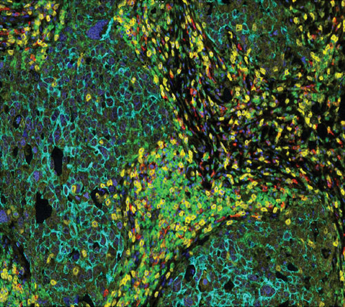
Pseudo composite of a breast cancer section labeled for killer T cells (CD8, yellow), helper T cells (CD4, green), B cells (CD20, red), epithelial cells (cytokeratin, aqua), and DAPI (blue). The imaging and analysis was accomplished with PerkinElmer’s Vectra imaging system and inForm software.
Quantitative Label-Free Drug Imaging
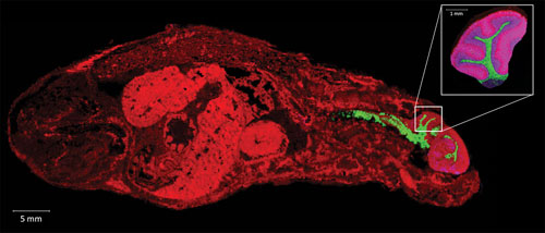
Mass spectrometry imaging (MSI) is used to visualize the spatial distribution of compounds, biomarkers, metabolites, peptides, or proteins by their molecular masses. MSI looks at whole-body sections to quantify molecules in all organs quickly, as compared to LC-MS/MS. It can be used on small organs where high resolution is needed, or dissection is difficult, such as the brain or eye tissues.
“MSI detects thousands of molecules from a simple microlayer of biological tissue, but the tissue is composed by biomolecules which interfere in the quantification process,” said Jonathan Stauber, Ph.D., CEO and CSO, ImaBiotech. “This is termed the biological matrix, or tissue, effect.”
“For example, if we detect one molecule corresponding to a drug at the same concentration in the liver and the brain, we have different signal intensities due to different biological-matrix compositions. This limited the possibility to quantify molecules.
“We have developed a quantification protocol and software, Quantinetix, which take into consideration the biological-matrix effect by normalizing the datasets and the images making absolute quantification possible,” said Dr. Stauber.
MALDI (matrix assisted laser desorption ionization) imaging detects compounds by using a laser that ionizes molecules, while LESA (liquid extraction surface analysis) uses a micropipette to extract and detect biomolecules at a selected position on the tissue section.
ImaBiotech’s services combine these two complementary technologies with Quantinetix software and a biomarker databank to provide images with quantification of drug candidates and markers of efficacy, or toxicity, in a single experiment. This approach allows pharmacokinetic and pharmacodynamics studies in whole-body tissue sections at a resolution of 15 microns.

Mass spectrometry imaging is used by ImaBiotech to reveal the distribution of drugs and metabolites, as in this whole-body section of a mouse.
Integrated Imaging Systems
Preclinical PET scanners with an integrated microCT have substantially improved the anatomical registration of PET predominately to the skeleton, yet little progress has been made in soft tissue contrast, even with the use of a CT contrast agent.
Integrated PET/MRI or SPECT/MRI systems offer many benefits. MRI uses no radiation, offers better soft tissue contrast, and provides molecular readouts. To date, preclinical PET imaging combined with MRI has been performed using two independent systems and a bespoke co-registration algorithm to fuse the images.
Mediso recently commercialized the first serially produced, fully integrated, automated PET/MRI system, the nanoScan PET/MRI, and a fully integrated, automated SPECT/MRI system, the nanoScan SPECT/MRI. Single systems enable use of the same imaging technology, imaging protocol, and biomarkers for small to large subjects.
According to Illes J. Muller, managing partner, preclinical PET/MRI and SPECT/MRI allow combination of radionuclide biomarkers with an MRI contrast agent on a routine basis, an attractive prospect for evaluating new drugs for oncology, neurology, and cardiovascular disease. Now, physiological/metabolic readouts can be combined with high-resolution, soft-tissue contrast as well as dynamic functional perfusion imaging.
In addition, SPECT provides the ability to perform multi-isotope imaging, probing two or more molecular pathways simultaneously by detecting isotopes with different emission energies, and has no physical limits in resolution. SPECT/MRI technology is less expensive. The labeling is easier, and no on-site cyclotron is required.
A potential major application for multimodal emission tomography combined with MRI is quantitative 3D imaging of tumor heterogeneity. To assess the spatial distribution of a given PET or SPECT biomarker within a tumor requires ultra-high resolution and high sensitivity and corrections for tumor perfusion. MRI is able to differentiate between healthy and dead tumor tissue for tumor response evaluation.
Expanding Companion Diagnostics
More and more, companion diagnostics, which are widely perceived in the field as in vitro tools, are relied upon to select the right patients for a targeted therapy, or to determine if the compound has brought about a therapeutic response.
“The companion diagnostic definition will gradually broaden to include the use of in vivo imaging agents,” said Karen Linder, Ph.D., research alliances manager at GE Healthcare. “Compounds labeled with a detectable tag such as 18F can be imaged noninvasively, and the distributions in the body can be followed over time after intravenous injection to provide a temporal three-dimensional picture of the distribution of the target in the body.”
“At its best, in vivo molecular imaging can allow selection of the right therapeutic for the right patient at the right time,” added Dr. Linder
In vivo imaging provides different information than in vitro diagnostics. In vitro blood tests may detect the presence of a shed antigen, circulating tumor cells, or other markers of interest, but information about the location of disease is not revealed. Location and extent of the disease affect treatment choices; use of an imaging agent that could sensitively, and specifically, identify recurrence could help determine between local or systemic therapy choices.
Furthermore, many in vitro diagnostic assays are typically performed on biopsy samples, and detect only what is present in the small sample. Accuracy can depend highly on when, and where, the biopsy was taken.
“For oncology applications, it is becoming more apparent that dramatic changes in tumor molecular markers can occur over time in response to clonal mutations, selective response to therapy, and the development of resistance. In vivo imaging tools that can help to demonstrate such changes are likely to be more widely appreciated as experience with such agents and clinical evidence grows,” concluded Dr. Linder.
High-Content Imaging Laboratories
The high-content imaging (HCI) laboratory provides expert information and guidance, and performs multiple imaging functions in a timely agreed-upon manner with key investigators.
“As an exclusive HCI laboratory, we perform the assay development, which may mean cloning and expression of cell lines specific for the project, optimize the cellular and manual or robotic operational aspects of the assay, execute the campaign, design and perform the image analysis, as well as the statistical and computational solutions for advancing the drug discovery efforts,” explained Ann F. Hoffman, principal scientist, Roche discovery technologies, and group leader, high content imaging, Hoffmann-La Roche.
For example, in analyzing for cellular cytotoxicity, a large net of imaging assays is cast to capture possible liabilities and then suggest additional in-depth studies that can be performed. With limited compound availabilities, the use of all reagents is then resourcefully relegated over a number of queries.
The results of all the data from the therapeutic platform panels, upon completion, are able to be put in perspective with respect to “gold standards,” known reference compounds, and historical data. This focused approach impacts the direction of compound progression and may lead to research findings not necessarily apparent.
As a better understanding of single cells and populations grows, such as stem and tumor cell differentiation and growth development, a way to link the results of HCI knowledge with the complicated and redundant signaling pathways and cellular processes will be required.
In the future, HCI laboratories will need to embrace and participate in the big data world that translational medicine and bioinformatics is establishing. The goal is to incorporate experimental research, preclinical HCI data, omics data, and next-generation-sequencing data for predictive analytical modeling that enables the forecasting of hypotheses and better clinical outcomes in the early stages of drug development.

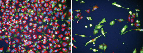
These images, generated by the high-content imaging group at Hoffmann-La Roche, display HeLa cells untreated (A) and treated (B) with a toxic compound. The phenotype of the toxic compound displays effects on cell morphology and structure as well as diminished cell numbers.


