January 15, 2009 (Vol. 29, No. 2)
Lloyd Dunlap
Usage Ranges from the Functional Analysis of Genes to the Diagnosis of Disease
PCR, although discovered over two decades ago, continues to serve as a key topic of interest at numerous meetings, underscoring its importance in molecular biology. Two upcoming conferences previewed in this article, CHI’s “Molecular Medicine Tri-Conference” and the “4th International qPCR,” will focus on PCR and its specific application to molecular diagnostics.
The technique uses a DNA polymerase to amplify or replicate DNA. As PCR progresses, the DNA generated is used as a template for further replication that sets a chain reaction in motion, generating millions of copies of the DNA piece. Real-time PCR is used to amplify and simultaneously quantify a targeted DNA molecule, adding immensely to the technique’s value in molecular biology in applications ranging from functional analysis of genes to the diagnosis of hereditary and infectious diseases.
Sean Ferree, Ph.D., research and development manager at NanoString Technologies, will highlight his company’s recently introduced nCounter® Analysis System.
“Using no enzymes or amplification, this novel system can analyze hundreds of genes in a single assay with high precision, even at low expression levels. We can assay directly from cell culture, tissue, or whole blood lysates,” he notes. “No sample purification is needed. We can also assay total purified RNA derived from formalin-fixed paraffin-embedded (FFPE) tissue samples.”
Currently, the system is being used in custom gene-expression studies for microarray follow-up, biomarker detection, and pathogen detection using mRNA. Off-the-shelf GPCR and kinase gene panels are under development. Dr. Ferree foresees that microRNA, copy-number variation, and direct protein analysis are additional future applications.
The NanoString system uses digital technology that is based on direct multiplexed measurement of gene expression and can detect attomolar concentrations at less than one copy per cell. Molecular barcodes composed of four fluorescent colors and six spots are used as reporters to analyze the expression levels of up to 550 genes simultaneously. Single molecule imaging is then used to detect and count hundreds of unique transcripts in a single reaction. Because digital detection doesn’t rely on relative levels of fluorescence, only the order of fluors on the barcode strings need to be detected.
Since the nCounter system requires no amplification of mRNA, the dynamic range necessary to measure the full range of expression in cells is simply the biological range of expression, typically three to four orders of magnitude, Dr. Ferree notes. Throughput is up to 72 samples in 24 hours, which at the 550-plex scale would correspond to 39,600 data points per day. The entire assay takes 18 hours for one 12-sample cartridge with the 12-hour hybridization step typically being accomplished overnight, followed by two hours on the liquid-handling prep station and four hours on the digital analyzer. Since the latter two steps are automated, the total hands-on time for the user is less than 15 minutes.
The system includes the automated prep station, a digital analyzer, the barcodes, and all reagents and consumables needed to perform the analysis.

Inhibition of myoblast fusion after TNF-alpha treatment
qPCR
Stephen Bustin, Ph.D., professor of molecular science at Barts and the London School of Medicine’s Institute of Cell and Molecular Science, will present at “qPCR2009.” Dr. Bustin is currently working on a real-time quantitative (qPCR) assay for the detection of Clostridium difficile, the bacterium responsible for a huge number of hospital-acquired infections. He expects that the assay will be more sensitive, more informative, and much less costly than current PCR-based assays.
Such assays, he notes, cost in the $20–$30 range per assay, and dedicated instrumentation is required. His goal is to develop a set of affordable tools that allow not just detection, but also a more detailed molecular characterization of C. difficile. Currently, he notes, there are many problems in the field of qPCR, and the problems are proving to be durable.
Dr. Bustin authored a paper citing such limitations in 2000, “but we haven’t progressed a lot since then,” he observes. In fact, “the field is littered with papers that are meaningless,” with the problems beginning with sample preparation, and extending to “how you normalize, analyze, and report results.”
“It is clear that a high percentage of publications utilizing qPCR technology, and especially those aiming to profile cellular RNA levels, report poorly designed, executed, and interpreted experiments and results,” Dr. Bustin states.
“Considerations of mRNA transcription, in vivo stability, regulation by miRNAs, tissue specificity of splice variants, allele-specific difference in expression, the lack of concordance between most mRNAs and their specified proteins, and the critical importance of post-translational modifications and questions of tissue heterogeneity all describe serious issues that are not being addressed in an adequate manner,” he concludes. “It will require a significant amount of courage, and a sea change in attitude from the research community to deal with this quagmire.”
On a positive note, there are several developments that Dr. Bustin finds encouraging. Among these he cites the introduction of less expensive, optimized reagents that make reaction assembly simpler and more consistent; the development of more intuitive analysis software to help with assay setup and project management; the introduction of advanced algorithms that allow more accurate quantitation; and the extension of the technology into novel areas such as high-throughput, nanoliter qPCR—specifically, microfluidic digital PCR, which is “an exciting new development that extends the scope of qPCR technology.”
Digital Applications
Ken Livak, Ph.D., senior scientific fellow at Fluidigm, explains the value of his company’s microfluidic chips for digital PCR and the system’s capability to analyze gene expression at the single cell level. Applications include cancer and stem cell research.
Noting that digital PCR has been around since the 1990s, but that it remains tedious and expensive, Dr. Livak says that microfluidics-based digital PCR makes the technique more sensitive and cost effective. The BioMark digital array functions as “the integrated circuit for biology,” Dr. Livak explains. “Reaction chambers within the chip are the size of a pencil dot, which contributes directly to the system’s stingy use of sample and reagents.”
The Fluidigm system has been used to identify and quantify ABL tyrosine kinase domain point mutations in samples from 28 patients. The digital array with its nanoscale channels, valves, and pumps partitioned samples into 12 panels, each panel containing 765 chambers. There is an improvement in detection of rare mutations as a consequence of partitioning before PCR, Dr. Livak observes.
He cites the example of a mixture containing a molecule of T3151 ABL in 100,000 molecules of unmutated ABL. If partitioned into 1,000 separate chambers, the chamber containing the single mutant molecule would only contain about 100 molecules of the unmutated ABL. This provides a 1,000-fold increase in relative concentration and allows for a theoretical 1,000-fold improvement in the detection sensitivity of PCR reactions.
Such sensitive detection of the T3151 mutation and other mutations may allow investigators to study the biology of clonal selection and evolution in the context of tyrosine kinase inhibitor therapy, resistance, and progression in chronic myelogenous leukemia, Dr. Livak concludes.
In addition, he notes, the BioMark system “lets you do higher- and higher- throughput analysis without a lot of robotics, making it more affordable so it can be used in more labs by more scientists.”
According to Fluidigm, microfluidics-based digital PCR makes the technique more sensitive and cost effective.
miRNA and Muscle Link
Also presenting at the upcoming conference in Germany will be Christian Thirion, Ph.D., CSO, Sirion Biotech. Dr. Thirion’s talk will focus on “Disregulated microRNAs—Novel Therapeutic Targets in Muscular Dystrophies.”
He notes that tumor necrosis factor alpha (TNF-α) is a prototypical inflammatory cytokine and a well-known mechanism for negating fusion in muscle cells. The injured muscles must be regenerated by myoblasts.
It is also well known that microRNAs are central regulators of muscle differentiation and that several cytokines might counter miRNA expression. In fact, Dr. Thirion points to 126 citations in the literature since 2005 linking miRNA and muscle.
Using a TaqMan low density array analysis, Dr. Thirion’s group found a number of miRNAs that were significantly upregulated and others that were significantly downregulated. When TNF-α was added, two key miRNAs in the previously upregulated group were downregulated. These were has-miR-133b and has-miR-206.
In addition TNF-a strongly upregulated miR-155, a microRNA linked to inflammation. When Agilent’s microarray analysis was substituted for TaqMan and TNF-α was added, all four downregulated miRNAs are muscle specific. Using the SC5 mouse cellular model, analysis confirmed that the effect of TNF-α is conserved on muscle-specific miRNAs across species.
From this work, the Sirion researchers concluded that TNF-α specifically acts on muscle-specific microRNA expression and that inhibition of muscle-specific microRNA expression is a potential disease mechanism in dystrophic muscle.
The group aims to further study the effects of cytokines/growth factors on microRNA expression, develop a functional analysis for regulated microRNAs, and intervene therapeutically on disregulated microRNA in muscle tissue to rescue non-fusion phenotype in vitro by microRNA over-expression. Finally, Dr. Thirion hopes to proceed to proof-of concept in an animal model for muscular dystrophy.
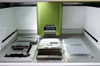
The nCounter Prep Station is the automated fluidic-handling component of the nCounter System. It processes samples post-hybridization to prepare them for data collection on the nCounter Digital Analyzer.

