January 15, 2009 (Vol. 29, No. 2)
Targeting Cell-Leucocyte Union Has the Potential to Establish New Therapeutics
Metastasis is the spread of cancer cells from their original site of origin in the primary tumor to distant tissues and organs. About 80% of tumors originate in the epithelial linings of the skin and various invaginations in the body such as the airways, milk ducts, and GI tract. Most of these are carcinomas (e.g., lung, breast, colon) and melanomas (melanocytes).
It is thought that the primary tumor develops from a single cell that has collected enough mutations to get its mitotic cycle stuck in the “on” position. Deregulated cell division results in a tumor consisting of ancestors from the original mutated cell. If cells continue to divide at a faster rate than they die, the tumor gets larger. But what causes some of these cells to metastasize, i.e., to gain the ability to leave their site of origin in the epithelium, migrate into the mesoderm, and disseminate throughout the body?
It is abundantly clear that metastasis is what makes cancer so deadly. If we just had to manage the primary tumor and not the spread, cancer survival would soar. The primary tumor can be surgically removed, the local area can be treated by radiation, and the patient can receive preventive chemotherapy. If spread hasn’t occurred, there is usually an excellent outcome. But once a cancer cell enters the vasculature or lymphatics and disseminates throughout the body, all the while continuing to divide, treatment is far more problematic.
Not only do the metastatic cancer cells migrate to virtually anywhere (lungs, brain, bone marrow, liver) but they are usually devilishly resistant to chemotherapy and radiation. Treatments to combat metastases often become palliative and no longer curative. Mortality occurs when vital organs fail due to tumor burden.
It is therefore surprising that so little is known about the onset of metastasis. While many of the molecules in the actual mechanics of metastasis are known, how it all gets started is still a mystery.
To my knowledge, the century-old theory of cancer cell fusion with tumor-associated leucocytes such as macrophages is really the only complete theory we have—potentially explaining most, if not all, aspects of metastasis and particularly its initiation
In this theory, metastasis is virtually a second disease imposed on the primary tumor cell. While the primary cell is notable for its deregulated cell cycle, it has little propensity to migrate away from its site of origin.
The fusion theory states that acquisition of a metastatic phenotype occurs when a healthy migratory leucocyte such as a macrophage fuses with a primary tumor cell. The resultant hybrid is a migratory cell with the uncontrolled cell division of the original cancer cell. A metastatic cell emerges, which, like a white blood cell, can migrate from the epithelium into the mesoderm, enter the circulatory system and travel to lymph nodes and distant organs.
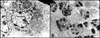
Autophagosomes, denoted by an asterisk (*), as seen through the electron microscope.
Lessons from Experimental Hybrids
I first encountered the fusion theory of metastasis in the early 1990s and was struck by its similarity to a well-known feature of evolutionary genetics—fusion of different cell types as a mechanism for creating new gene-expression patterns and biodiversity.
With this large biological precedent as a stimulus, my research group created experimental hybrids in the laboratory between normal macrophages ultimatelyand a mouse melanoma cell line that was only weakly metastatic in mice. We were amazed to find that many of the hybrid clones were now markedly metastatic when implanted into mice.
This finding led us to a now 15-year study of the fusion theory. We have studied experimental macrophage-melanoma hybrids from many angles—chromosome content, genes expressed that are associated with metastasis, capability for chemotactic migration, and many other features. We were looking for similarities of hybrids to metastatic melanoma in humans. The thought was that the extent to which experimental macrophage-melanoma hybrids might resemble metastatic melanoma cells in humans could provide support and direction for the theory, though not ultimate proof.
We established a long list of molecules expressed by macrophage-melanoma hybrids that were also characteristic of metastatic melanoma cells and normal macrophages alike. Thus this “quacks like a duck” approach gave us some confidence that we might be on the right track—we could create a phenotype similar to that of authentic metastatic melanoma cells by fusing normal macrophages with non-metastatic melanoma cells.
But, not only did the experimental hybrids express a number of metastasis-associated proteins, they also expressed macrophage-like glycosylation structures associated with migration and metastasis. This involved a leukocytic glycosylation system where complex sugar structures known as β1,6-branched oligosaccharides are attached to N-glycoproteins, thereby orienting the cell toward a migratory phenotype.
A rate-limiting enzyme in this system is N-acetylglucosaminyltransferase V (GnT-V), a Golgi complex enzyme that is highly expressed in macrophages and other migratory leucocytes and also widely associated with metastasis.
GnT-V and its enzymatic products, b1,6-branched oligosaccharides conjugated to N-glycoproteins, are associated with poor outcome in melanoma and carcinomas of the breast, colon, lung, GI tract, and endometrium.
GnT-V is a promiscuous enzyme with many N-glycoprotein substrates such as motility-associated integrins, adhesion molecules, and growth factor receptors. As such, GnT-V is a master regulator of migration and metastasis, initiating cascade reactions by activating numerous protein substrates through altered glycosylation. Our results with macrophage-melanoma hybrids could explain why so many cancers express GnT-V and β1,6-branched oligosaccharides while their normal cell counterparts do not.
Autophagy: Energy Independence?
We were surprised to find that experimental hybrids and metastatic melanoma cells expressing high levels of β1,6-branched oligosaccharides also seemed to be in a state of autophagy.
Autophagy is “self eating” wherein a cell’s cytoplasmic components are engulfed and digested as a temporary energy source in times of nutrient deprivation, for example under hypoxic conditions in the absence of a blood supply. While autophagy is associated with cancer cell death pathways, paradoxically it is also a predictor of worse patient outcome in a variety of cancers.
We proposed that, should autophagy be linked to phagocytosis in cancer cells as it is in macrophages, nutrients could be continuously phagocytosed from external sources (cellular debris, matrix fragments, etc.) and digested in autophagolysosomes. Perhaps such a system would ensure a positive metabolic energy balance during migration from avascular areas to more vascularized ones or during tumor neo-angiogenesis.
New Targets for Therapy
Thus, while much more work needs to be done to establish whether the fusion theory is operative in human cancer, circumstantial evidence is growing for the widespread involvement of fusion in tumor progression.
In my opinion, a stepwise mutation model cannot readily explain acquisition of the complex gene-expression patterns necessary for migration and metastasis. Not only does the fusion theory explain the onset of metastasis through a single initiating event but actual data from macrophages-melanoma hybrids reveal that fusion causes just what the theory predicts it should.
Looking ahead, this could open up many new potential targets for therapy, for example, development of therapies for suppression of cancer cell fusion or for targeting GnT-V, β1,6-branched oligosaccharides and autophagy. But, we need to learn more before such ideas can be translated to the clinic. I would urge more academic scientists and biotechology companies to consider entering this most interesting area of cancer research.
John M. Paweleck, Ph.D. ([email protected]), is research affiliate in the department of dermatology at the Yale Cancer Center.

